How to Read a UC Colonoscopy Report (Erythema, Friability, Ulcers)
By the Aidy Editorial Team
First Published Nov 23, 2025Last Updated Jul 8, 2026
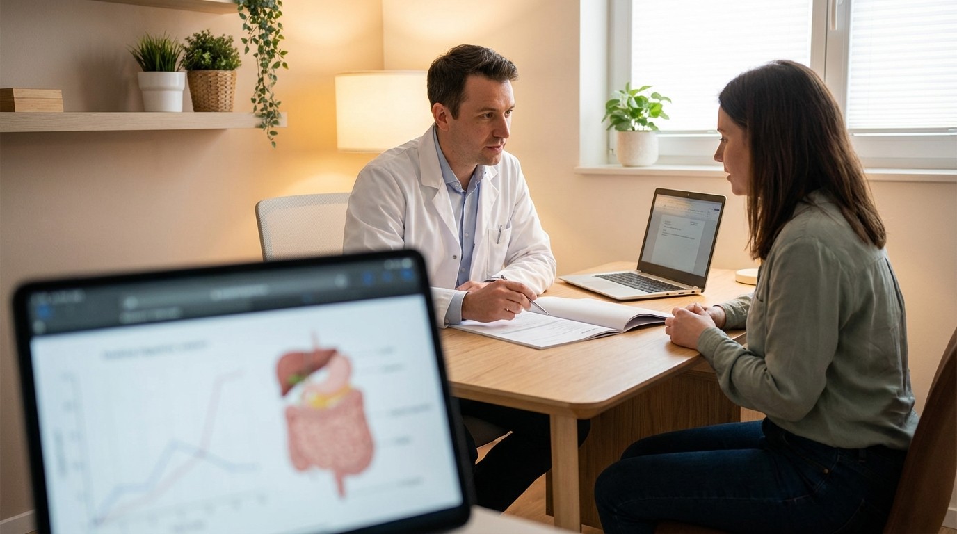
A colonoscopy report can feel like a mix of shorthand and medical vocabulary. For ulcerative colitis (UC), it is mainly a snapshot of what the lining of the colon and rectum looked like during the exam, plus any photos, a severity score (sometimes), and a plan for follow-up. Understanding a few common terms can make it easier to track patterns over time and to know what to ask about while waiting for biopsy results and next-step recommendations.
What the endoscopist saw: common UC terms and severity scores
In an ulcerative colitis colonoscopy report, the “Findings” section often describes what the surface of the colon looked like and how easily it bled. Three terms show up often:
Erythema (erythema colonoscopy): redness of the lining, a common sign of irritation or inflammation.
Friability (friability meaning colonoscopy): tissue that bleeds easily when touched or with scope movement, which can point to more active inflammation.
Ulcerations (ulcerations UC): breaks in the lining. Reports may also mention erosions (smaller, shallow defects) versus ulcers (larger, deeper sores).
Many reports include an endoscopic score to summarize severity. A common one is the Mayo endoscopic subscore , which runs from 0 to 3. In simple terms, Mayo 1 is often described with mild redness and mild friability, Mayo 2 adds more obvious inflammation and erosions, and Mayo 3 includes ulceration and spontaneous bleeding. Some reports also mention “vascular pattern,” meaning whether normal-looking blood vessels can be seen (inflammation can make that pattern fade or disappear). [1]
Another scoring system sometimes used is the Ulcerative Colitis Endoscopic Index of Severity (UCEIS). It scores three features, vascular pattern, bleeding, and erosions/ulcers , each with set definitions, and totals them into a number (higher generally means more inflammation). [2]
People may also see the phrase endoscopic remission UC or mucosal healing in follow-up notes. Patient organizations commonly describe mucosal healing as no active disease seen on colonoscopy, such as no ulcers and no bleeding. [3]
What the lab found: biopsies, pathology, and next steps to discuss
Even when the lining looks improved, gastroenterologists often take small tissue samples (biopsies). Those samples are reviewed under a microscope and reported separately as biopsy results UC or “pathology.” A helpful way to read this part is to look for three categories of information: (1) whether inflammation is still present under the surface, (2) whether there are signs of infection or other causes of inflammation, and (3) whether there are any precancer changes (often described as “dysplasia”). The colonoscopy report may list how many samples were taken and from which areas.
Timing can reduce anxiety. The endoscopist can usually share initial visual findings right after the procedure, but pathology results often return later , and final recommendations may depend on those results. Many health systems note that biopsy processing commonly takes about 1 to 2 weeks , depending on the lab and whether extra testing is needed. [4]
Follow-up planning often includes when the next colonoscopy should happen. For people with long-standing colonic inflammatory bowel disease (including UC), professional guidance generally recommends starting screening for dysplasia 8 to 10 years after diagnosis (earlier in some higher-risk situations) and then repeating surveillance about every 1 to 5 years based on individual risk factors and prior exam quality. [5]
Questions that can help guide the next discussion include: Which score was used (Mayo or UCEIS), what was the most inflamed area, whether friability or ulcers were seen, when biopsy results are expected, and what factors drove the recommended next colonoscopy interval.
References
This article is for educational purposes and is not medical advice. It is researched against current AGA clinical guidelines and peer-reviewed sources. Always discuss treatment decisions with your care team.
Understanding Your UC Lab Results: CRP, Calprotectin, and More ›